In lectures that featured dozens of surgical videos, Robert Spetzler, MD, one of the world’s great surgeons, on Wednesday showed fellow surgeons and medical students how one gets from here to there in the brain, when “there” is a place where few dare to go.
“Neurosurgery is all about anatomy,” said Dr. Spetzler, Director of the Barrow Neurological Institute and the J.N. Harber Chairman of Neurological Surgery at the University of Arizona. “All the corridors I describe are based on anatomy.”
Even difficult lesions or malformations are accessible through safe entry zones, he said. “There are multiple routes available. And with appropriate planning, it is our job as neurosurgeons to find the right route to these lesions.”
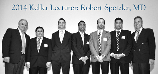
Dr. Spetzler spoke as the 2014 Keller Lecturer in Surgical Neuroanatomy & Research. The third annual Keller Lectureship was hosted by the Mayfield Education & Research Foundation and the University of Cincinnati Department of Neurosurgery on the UC College of Medicine campus. Additional support came from Stryker®. The lecture is named for Jeffrey T. Keller, PhD, Research Professor of Neurosurgery & Anatomy and Cell Biology at UC and the Mayfield Clinic.
Dr. Spetzler’s two-part lecture was titled, “Traversing the Brainstem and Third Ventricle.”
Those who attended the Lectureship witnessed the work of this remarkable surgeon in riveted silence. In case after case, Dr. Spetzler deftly combined state-of-the-art instrumentation and image-guidance technology with steady hands and “a little bit of patience” to reach and extract life-threatening cavernous malformations of the brainstem and the adjoining spinal cord. These vascular malformations afflict young and old alike, are prone to bleeding, and can grow quite large.
Dr. Spetzler said that cavernous malformations “have taught us more than anything about how to get to deep areas of the brain.”
Dr. Spetzler worked almost exclusively without retractors, surgical instruments that hold a wound open or grasp tissue that might otherwise obstruct the surgeon’s view. In their place Dr. Spetzler used the patient’s own anatomy, his own alignment with the patient’s head, and in some cases gravity. “I’m not against retractors,” he said, “I just make minimal use of them.”
He worked deliberately and stressed the importance of patience. “I spend a lot of time at the end of the procedure taking out the last pieces of the cavernous malformation,” he said. “To prevent recurrence I take out anything with little white fibers, any potential residuals. I take my time to get every bit that is accessible.”
A world-renowned neurosurgeon who specializes in cerebrovascular disease and skull base tumors, Dr. Spetzler has co-edited a number of textbooks, including the Color Atlas of Microneurosurgery.
George Mandybur, MD, a Mayfield neurosurgeon who attended the lectures, recalled seeing Dr. Spetzler operate during a rotation at Barrow during his senior year of medical school. “I remember one of his cases in particular,” Dr. Mandybur recalled. “A high-end businessman who had a brainstem cavernous malformation flew in on his private jet. I saw the MRI of this lesion in the middle of the brainstem and thought, ‘That’s got to be no man’s land.’ And he goes after it and takes it out and the patient does pretty well. It was very impressive.”
Despite risks associated with removing cavernous malformations in the brainstem and spinal cord, ignoring a malformation that is causing symptoms portends a poor outcome for the patient.
Barrow Ruptured Aneurysm Trial
In an impromptu addition that addressed specific interests of UC neurosurgery residents, Dr. Spetzler presented recent research on aneurysm treatments, including the six-year Barrow Ruptured Aneurysm Trial (BRAT). The trial compared the results of more than 400 patients whose aneurysms had either been clipped or coiled by one of four experienced Barrow surgeons. Although national and international trends continue in the direction of minimally invasive endovascular coiling, the BRAT trial illustrated distinct benefits of clipping, which involves opening the skull.
After 6 years, patients in the clipping group had 96 percent obliteration of their aneurysm, compared to 48 percent in the coiling group. Retreatments were required for 13 percent of patients in the coiling group at 6 years but only 4 percent in the clipping group.
“BRAT was a single-institution trial, and there is an absence of conclusive data to determine which treatment is better,” Dr. Spetzler said. “The risk of rebleeding and retreatment favors clipping. But we need another large randomized trial, as hard as that would be.”
See the published 3-year results of BRAT >>
Resident research presentations
Five specially selected neurosurgery residents gave research presentations, which were judged by Dr. Spetzler; John. M. Tew, MD, Mayfield neurosurgeon and Professor of Neurosurgery, Radiology and Surgery; and Kerry Crone, MD, Residency Director and Professor of Pediatric Neurosurgery. The residents placed as follows:
1st Place: Optic Nerve Surface Temperature During Intradural Anterior Clinoidectomy: A Comparison between High-speed Diamond Burr and Ultrasonic Bone Curette
Varun R. Kshettry, MD, Cleveland Clinic
2nd Place: A Comparison of Operative Exposure between the Le Fort I Osteotomy and the Expanded Endoscopic Endonasal Approach to the Clivus
Christopher Sanders Taylor, MD, University of Cincinnati
3rd Place: The Medial Corridor to the Medial/Posteromedial Cavernous Sinus
Almaz Kurbanov, MD, University of Cincinnati
Honorable Mention: Minipterional Approach: Anatomical Considerations, Clinical Applications, and Limitations
Mohab Mohamed Nageeb Darwish, MD, Drexel University
Honorable Mention: Microsurgical and Endoscopic Anatomy of the Retrosigmoid Inframeatal Approach
Roberto Colasanti, MD, Ohio State University
Previous Keller Lecturers
2013 Takeshi Kawase, MD, PhD, Keio University, Tokyo >>
2012 Albert L. Rhoton, Jr., MD, University of Florida >>
— Cindy Starr